
Cyclotron in Thunder Bay Saves Lives
THUNDER BAY – Healthbeat – Having a cyclotron just minutes, rather than hours, away is good news for both patients and healthcare research in Northwestern Ontario.
Your donation to the Exceptional Cancer Care Campaign will help fund important local cutting-edge research that will help develop tomorrow’s cancer treatments. Please give generously at healthsciencesfoundation.ca/ECC or call (807) 345-HOPE (4673).
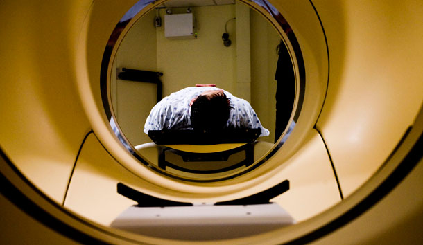
The cyclotron produces fluorine-18, the material that is injected into patients prior to a PET-CT scan, which enables healthcare professionals to detect cancer, to diagnose and to prepare a care path. Until now, Thunder Bay Regional Health Sciences Centre (TBRHSC) has had to purchase this material from Southern Ontario.
Having a cyclotron on the TBRHSC campus will provide a more ready and reliable access to these isotopes, particularly for patients waiting for PET-CT scans. Although it has rarely happened, airline pilots can refuse to fly with radioactive material. Weather can also cancel or delay flights. In those cases, nuclear medicine staff might be forced to cancel or postpone some patients’ appointments.
Also, the cost involved in transporting these isotopes is further magnified because of their relatively short half-lives. The half-life of FDG (fluorodeoxyglucose) is about 110 minutes and currently must travel to Pearson International Airport in Toronto before the two-hour flight to Thunder Bay. “By the time it reaches Thunder Bay, only about 1/8 of the material is remaining,” says Michael Campbell, Director of Research Operations at Thunder Bay Regional Research Institute (TBRRI) and Cyclotron Project Manager. “So to treat four patients, which we typically see in a day, TBRHSC actually needs to purchase enough material to treat 32 patients.”
The Cyclotron and Radiopharmacy will be housed in the new Health Services Centre, being built adjacent to TBRHSC and will produce fluorine-18 for use in imaging tests at TBRHSC. The cyclotron will also provide isotopes for medical imaging research and clinical trials at TBRRI. Of particular interest is the potential of medical imaging to help select the most appropriate candidates for certain clinical trials and cancer treatments. “FDG answers the question ‘Is there cancer present?’” says Campbell. “But it doesn’t answer the question ‘How should we treat it?’ Through clinical trials using a combination of FDG, new tracers and other isotopes with longer half-lives, clinicians can have better information on what drug to use in treatment so they get the patient on the right drug the first time, saving the patient valuable time as well as unpleasant and unnecessary side effects.
TBRRI scientist and Adjunct Professor at Lakehead University and Northern Ontario School of Medicine, Dr. Chris Phenix and his team are already working to develop PET as a method to indicate a cancer’s potential to spread or resist treatment and plan the most appropriate, personalized treatment for a patient.


